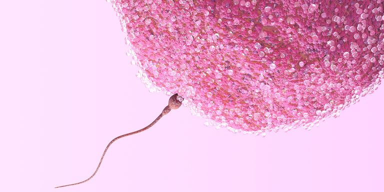
Infertilità causata da esposizioni al 4G (LTE)
Le esposizioni prolungate a microonde della tecnologia 4G, sopprimono la fertilità nei ratti di sesso maschile.
Col 5g accadrà a livello di fertilità, lo stesso identico fenomeno.
Le esposizioni prolungate a microonde della tecnologia 4G, sopprimono la fertilità nei ratti di sesso maschile.
Col 5g accadrà a livello di fertilità, lo stesso identico fenomeno.
12 luglio 2016 – “www.casalenews.it”
Chi era presente venerdì scorso all’Auditorium Santa Chiara per l’incontro su “Wifi, cellulari & co” ha capito che ‘indossare’ un telefonino (perché tale è l’intimità raggiunta con questo strumento di comunicazione) può provocare danni alla salute, anche se il mondo scientifico non lo ha ancora dimostrato.
Nell’incontro organizzato da Mamme in cerchio, Albero di Valentina e Passi di vita Onlus, avente per oggetto proprio l’informazione sui rischi provocati dalla prolungata esposizione a onde elettromagnetiche, autorevoli esperti si sono espressi e confrontati sull’entità e sulla peculiarità delle conseguenze di tale esposizione.
Ha aperto la serata l’epidemiologo Corrado Magnani, che ha richiamato i risultati degli studi scientifici ufficiali, per i quali non ci sono evidenze di una pericolosità delle onde elettromagnetiche ai livelli di esposizione attualmente consentiti, benché le correlazioni con gravi malattie siano in fase di approfondimento.
Terminato il suo intervento, si è voltata pagina: il dottor Orio, vice-presidente dell’Associazione Elettrosensibili, ha riconosciuto i risultati divulgati dalla ricerca scientifica, ma ha dato conto di evidenze medico-sanitarie rilevanti: a fronte della diffusione rapidissima di cellulari e tecnologie wi-fi, i danni al sistema neurologico di bambini e adulti sono aumentati, e con loro disturbi all’apparato riproduttivo maschile.
Eloquenti le immagini mostrate: le parti a contatto con il telefonino subiscono l’aggressione di onde elettromagnetiche in una forma inedita fino ad oggi.
Sono poi emersi altri aspetti – oggetto di studi indipendenti – che rafforzano l’idea di dover essere prudenti quando ci affidiamo alla connessione wireless, perché, come il professor Magnani ha suggerito, si tratta di una tecnologia entrata nelle nostre case prima di averne sperimentato e ‘pesato’ gli effetti.
Effetti nefasti, li conoscono bene gli elettrosensibili: persone che – con intensità diverse – registrano l’impossibilità di maneggiare apparecchi come cellulari, computer, microonde.
Alcuni erano presenti all’incontro e hanno portato testimonianze toccanti. È verosimile aspettarsi un aumento di elettrosensibili, e ci si augura che la tecnologia migliori la schermatura degli apparecchi e che la vocazione per il wi-fi venga soppiantata da sistemi più inequivocabilmente sicuri per la salute.
Una serie di consigli dispensati dal professor Sergio Crippa ha, a questo proposito, semplificato la materia: accorgimenti casalinghi praticabili e utili.
Il materiale raccolto dalle associazioni promotrici, che hanno avuto il sostegno e il patrocinio del Comune di Casale e dell’Ordine dei Farmacisti di Alessandria, per la serietà con cui è stata impostata la serata e per la capacità di coinvolgere personalità di prim’ordine, è a disposizione di chi volesse approfondire il tema e per chi – in attesa di evidenze scientifiche (per le quali occorre tempo, quale che sia l’esito) – volesse divulgare l’invito alla prudenza nell’utilizzo del wi-fi. Internet e cellulari si’, ma non a discapito della salute.
Associazioni Mamme in Cerchio – L’Albero di Valentina – Passi di vita onlus
Fonte:
Auditorium Santa Chiara
via F. Cane, 31 – Casale Monferrato (AL)
Serata informativa sull’uso consapevole di cellulari, wifi, ecc.,
e i rischi potenziali per la salute.
Organizzata dalle associazioni
Mamme in Cerchio, L’Albero di Valentina, Passi di Vita Onlus,
con il patrocinio
del Comune di Casale Monferrato e dell’Ordine dei Farmacisti della provincia di Alessandria.
[Per maggiori dettagli, vedere la locandina qui sotto.
Cliccare sulla immagine per ingrandirla.]

Una presentazione della serata su “Il Monferrato”, di Marco Bertoncini.
[Cliccare sulla immagine per ingrandirla.]
[EN]
This US Navy report from 1972 documents the connection of 122 bioeffects to microwaves. Dr. Zorach Glaser, PhD of the US Navy presented a research report covering more than 2,200 studies, which link weak wireless signals (microwave radiation) to more than 122 biological effects. “Bibliography of Reported Biological Phenomena (Effects) and Clinical Manifestations attributed to Microwave and Radio-Frequency Radiation.”
![]()
[IT]
Questo rapporto della Marina degli Stati Uniti del 1972 documenta il nesso tra 122 bioeffetti e le microonde.
Il Dr. Zorach Glaser, PhD della Marina degli Stati Uniti ha presentato un rapporto di ricerca che copre più di 2.200 studi, i quali collegano i segnali wireless deboli (radiazione a microonde) a più di 122 effetti biologici.
“Bibliografia dei Fenomeni Biologici Riferiti (Effetti) e delle Manifestazioni Cliniche attribuiti alla Radiazione in Microonde e Radiofrequenza.”
Click on the picture below to access the PDF file of the research report/Cliccare sulla immagine sottostate per accedere al file PDF del rapporto:
Source/Fonte: https://apps.dtic.mil/sti/citations/AD0750271
 Realizzata con il patrocinio dell’Ordine dei Farmacisti della Provincia di Lecce dal Comitato Lecce Via Cavo, insieme alle Associazioni Consumatori Codacons e Codici, a Csv Salento e all’Associazione Italiana Elettrosensibili, questa locandina nasce dalla necessità di informare la popolazione sui possibili rischi per la salute derivanti dall’uso di cellulari, smartphone, tablet, telefoni cordless, apparecchiature Wi-Fi e Wireless in genere, anche per livelli di esposizione alle Radiofrequenze/Microonde inferiori ai limiti di legge.
Le Radiofrequenze/Microonde, utilizzate dalle apparecchiature Wireless, sono state classificate nel 2011 come “possibile cancerogeno per l’uomo” dall’Agenzia Internazionale per la Ricerca sul Cancro (I.A.R.C.), afferente all’Organizzazione Mondiale della Sanità (O.M.S.).
Da più parti è stato invocato il Principio di Precauzione, secondo il quale, in attesa di ulteriori informazioni, è importante adottare misure pratiche per ridurre l’esposizione a queste fonti di inquinamento ambientale.
Numerose sono le evidenze scientifiche (vd. la nostra sezione Riferimenti scientifici, dove ne viene riportata una parte) che correlano l’esposizione alle Radiofrequenze/Microonde con: danni al DNA, insorgenza di tumori maligni al cervello, malattie neurologiche e neurodegenerative, alterazioni del ritmo cardiaco, deficit di apprendimento e di memoria, disturbi cognitivi, mal di testa, insonnia, sintomi di Elettrosensibilità, infertilità, diminuzione dell’udito e tinnito, autismo nei bambini e molto altro.
Sono tra l’altro recentemente stati pubblicati i primi risultati di un importante studio condotto dall’ente governativo statunitense National Toxicology Program, costato finora ben 25 milioni di dollari, dai quali si evince un incremento significativo di due tipi di cancro nei ratti esposti alle Radiofrequenze/Microonde: Glioma (tumore della glia del cervello) e Schwannoma (tumore maligno del cuore).
I bambini andrebbero maggiormente tutelati, perché sono particolarmente vulnerabili agli effetti dannosi delle Radiofrequenze in quanto la loro gracile costituzione e la loro esposizione precoce determinano un rischio maggiore di manifestare tumori e malattie neurodegenerative in età adulta.
La locandina sarà diffusa nelle farmacie di Lecce e provincia, in altri esercizi commerciali e negli studi medici.
Chiunque può contribuire alla sua diffusione scaricando e stampando il file in alta qualità il cui link trovate in calce alla pagina.
Il formato consente una stampa ottimale anche di locandine di grandi dimensioni (A3-A2).
14 giugno 2016 – “www.terranuova.it”
Con il patrocinio dell’Ordine dei Farmacisti della Provincia di Lecce, il Comitato Lecce Via Cavo, insieme alle Associazioni Consumatori Codacons e Codici, a Csv Salento e all’Associazione Italiana Elettrosensibili, ha realizzato la locandina “Dai voce al tuo cervello”.

«L’iniziativa nasce dalla necessità di informare la popolazione sui possibili danni alla salute legati all’uso di cellulari, cordless, tablet, wifi, etc. – spiegano i promotori – Le radiofrequenze, utilizzate dalla tecnologia senza fili sono state classificate nel 2011 dall’Agenzia Internazionale per la Ricerca sul Cancro (IARC), afferente all’Organizzazione Mondiale per la Salute (OMS), come “possibile cancerogeno per l’uomo”. In quell’occasione il direttore della IARC ha affermato che “in attesa di ulteriori informazioni è importante adottare misure pratiche per ridurre l’esposizione, per esempio usando gli auricolari e i messaggi non in voce”. Da allora sono emerse nuove evidenze scientifiche che correlano l’esposizione alle radiofrequenze con l’insorgenza di tumori maligni al cervello, disordini neurologici, danni al Dna, diminuzione dell’udito, sintomi di elettrosensibilità, alterazioni del ritmo cardiaco, insonnia, cali di memoria, disturbi cognitivi, etc.».
«La locandina, che sarà diffusa nelle farmacie di Lecce e provincia, ed anche in altri esercizi commerciali e studi medici, suggerisce alcuni comportamenti per ridurre la propria esposizione alle radiofrequenze, come quello di tenere lontano il cellulare quando si aspetta la connessione o mentre si invia un sms, o spegnere il wifi quando non è in uso, soprattutto durante la notte per evitare disturbi del sonno, staccando la spina. I bambini andrebbero maggiormente tutelati, poiché sono particolarmente vulnerabili alle radiofrequenze e per la loro esposizione precoce corrono un rischio maggiore di manifestare in età adulta malattie neurodegenerative e tumori. L’Ordine dei Farmacisti della Provincia di Lecce, da sempre sensibile e attento alle problematiche sanitarie ricopre ancora una volta un ruolo di primo piano nella tutela della salute pubblica e della prevenzione delle malattie. L’uso del cellulare e dei dispositivi wifi è diffusissimo ma pochi conoscono i reali danni alla salute che ne potrebbero derivare, anche per livelli di esposizione inferiori ai limiti di legge. Tantissimi studi sono stati realizzati dal 2011 in poi. Ultimo in ordine di tempo è quello condotto dall’ente governativo statunitense National Toxicology Program e costato fino ad ora 25 milioni di dollari. I primi risultati, pubblicati qualche giorno fa, hanno evidenziato un incremento significativo di due tipi di cancro nei ratti esposti alle radiofrequenze: glioma (tumore delle cellulari gliali del cervello) e schwannoma maligno del cuore. Anche uno studio recente condotto dall’Istituto Ramazzini di Bologna ha evidenziato un aumento significativo di schwannoma maligno del cuore nei ratti esposti a campi elettromagnetici di bassissima frequenza. Il professor David Carpenter, direttore dell’Istituto per la Salute e l’Ambiente dell’Università di Albany (New York) ha affermato: “Siamo a un punto di svolta. Sarà molto difficile per i negazionisti non riconoscere l’associazione tra radiofrequenze e tumori”».

Fonte:
This is the presentation I gave to scientists at NIEHS in Triangle Park, North Carolina, May 9, 2016. Title of the talk is, “Electrosmog, the missing link as it relates to cancer, reproductive problems and electrohypersensitivity.”
[Effetti del Wi-Fi a 2,45 GHz sulla fertilità dei ratti maschi.
La versione PDF completa dell’articolo è scaricabile dal link in calce alla pagina.]
Electromagn Biol Med. 2015 Mar;34(1):37-42. doi: 10.3109/15368378.2013.869752. Epub 2014 Jan 24.
By:
Dasdag S1, Taş M, Akdag MZ, Yegin K.
1Department of Biophysics, Faculty of Medicine, University of Dicle , Diyarbakir , Turkey.
Article history
Received: 3 May 2013
Revised: 15 November 2013
Accepted: 17 November 2013
Published online: 21 January 2014
Keywords
2.4 GHz Wi-Fi; electromagnetic fields; long-term exposure; radiofrequency; reproduction; testes
The aim of this study was to investigate long-term effects of radiofrequency radiation (RFR) emitted from a Wireless Fidelity (Wi-Fi) system on testes. The study was carried out on 16 Wistar Albino adult male rats by dividing them into two groups such as sham (n: 8) and exposure (n: 8). Rats in the exposure group were exposed to 2.4 GHz RFR radiation for 24 h/d during 12 months (1 year). The same procedure was applied to the rats in the sham control group except the Wi-Fi system was turned off. Immediately after the last exposure, rats were sacrificed and reproductive organs were removed. Motility (%), concentration (×10(6)/mL), tail defects (%), head defects (%) and total morphologic defects (%) of sperms and weight of testes (g), left epididymis (g), prostate (g), seminal vesicles (g) were determined. Seminiferous tubules diameter (μm) and tunica albuginea thickness (μm) were also measured. However, the results were evaluated by using Johnsen’s score. Head defects increased in the exposure group (p < 0.05) while weight of the epididymis and seminal vesicles, seminiferous tubules diameter and tunica albuginea thickness were decreased in the exposure group (p < 0.01, p < 0.001, p < 0.0001). However, other alterations of other parameters were not found significant (p > 0.05). In conclusion, we observed that long-term exposure of 2.4 GHz RF emitted from Wi-Fi (2420 μW/kg, 1 g average) affects some of the reproductive parameters of male rats. We suggest Wi-Fi users to avoid long-term exposure of RF emissions from Wi-Fi equipment.
Source/Fonte:
http://www.ncbi.nlm.nih.gov/pubmed/24460421
Versione PDF completa scaricabile al seguente link:
[Effetti del Wi-Fi a 2,45 GHz sulla fertilità dei ratti maschi.]
Cell J. 2015 Summer; 17(2): 322–331. PMCID: PMC4503846
By:
Saeed Shokri, Ph.D,1,* Aiob Soltani, M.Sc,2 Mahsa Kazemi, M.Sc,3 Dariush Sardari, Ph.D,2 and Farshid Babapoor Mofrad, Ph.D2
Article history
Received: 24 April 2014
Accepted: 18 September 2014
Published online: 11 July 2015
Keywords
Apoptosis; Electromagnetic Radiation; Testis; Spermatogenesis
In today’s world, 2.45-GHz radio-frequency radiation (RFR) from industrial, scientific, medical, military and domestic applications is the main part of indoor-outdoor electromagnetic field exposure. Long-term effects of 2.45-GHz Wi-Fi radiation on male reproductive system was not known completely. Therefore, this study aimed to investigate the major cause of male infertility during short- and long-term exposure of Wi-Fi radiation.
This is an animal experimental study, which was conducted in the Department of Anatomical Sciences, Faculty of Medicine, Zanjan University of Medical Sciences, Zanjan, IRAN, from June to August 2014. Three-month-old male Wistar rats (n=27) were exposed to the 2.45 GHz radiation in a chamber with two Wi-Fi antennas on opposite walls. Animals were divided into the three following groups: I. control group (n=9) including healthy animals without any exposure to the antenna, II. 1-hour group (n=9) exposed to the 2.45 GHz Wi-Fi radiation for 1 hour per day during two months and III.7-hour group (n=9) exposed to the 2.45 GHz Wi-Fi radiation for 7 hours per day during 2 months. Sperm parameters, caspase-3 concentrations, histomorphometric changes of testis in addition to the apoptotic indexes were evaluated in the exposed and control animals.
Both 1-hour and 7-hour groups showed a decrease in sperm parameters in a time dependent pattern. In parallel, the number of apoptosis-positive cells and caspase-3 activity increased in the seminiferous tubules of exposed rats. The seminal vesicle weight reduced significantly in both1-hour or 7-hour groups in comparison to the control group.
Regarding to the progressive privilege of 2.45 GHz wireless networks in our environment, we concluded that there should be a major concern regarding the timedependent exposure of whole-body to the higher frequencies of Wi-Fi networks existing in the vicinity of our living places.
Electromagnetic radiation (EMR) from different sources, such as microwave ovens, radar, satellite links, wireless communication, frequency modulation (FM) radio and television (TV) transmitters/ antennas, is the main part of indoor- outdoor electromagnetic field exposure spectrum (1, 2). Widespread usage of industrial, scientific, medical, military and domestic applications with 2.45-GHz radio-frequency radiation is inevitable in today’s world. As the Wi-Fi technology is low cost and operates in the unlicensed spectrum at 2.40-2.4 GHz, the leakage of Wi-Fi radiation into the environment is unavoidable (3, 4).
It has been suggested that male infertility during the past several decades is related to the direct or indirect exposure to certain environmental factors such as radio-frequency electromagnetic waves (RF-EMW) (5, 6). The effects of 2.45-GHz EMR on reproductive system have already been shown (7–10). Kumar et al. (11) showed 2.45 GHz microwave exposure causes an increase in caspase-3 and creatine kinase activities in the sperm in addition to a decrease in plasma levels of testosterone and melatonin in the exposed rat. In vitrostudy by Avendano et al. (12), focused on the effect of Wi-Fi radiation on the motility reduction and DNA fragmentation of human spermatozoa. The negative effect of Wi-Fi emitting RF-EMW has been also reported on the ex vivo human sperm parameters (13), sexual behavior (14) and testis structure of exposed animals (15). It is believed that exposure to EMR can enhance production of reactive oxygen species (ROS) (9, 12,15–18). An increase in lipid peroxidation levels in addition to a decrease in antioxidant enzymes and vitamin A and E levels (11, 19) can explain some aspects of 2.45-GHz EMR effect on reproductive tissues of male rats. Kim et al. (20) showed that the effect of exposure to 2.45-GHz EMR on proliferation and differentiation of spermatogonia is correlated with serum sex hormone level. In parallel with defect in spermatogenesis process, the negative effects of 2.45-GHz EMR on histopathological changes and apoptosis status of rat testis are inevitable (7). Nowadays 2.45 GHz wireless networks have become much more commonplace in our environment (21). Wireless devices have been widespread used in our living and working environments for longer exposure times than wireless phones which may have an untoward influence on health (2). According to the Bioinitiative Report (http://www.bioinitiative.org/), current safety guidelines for electromagnetic field (EMF) exposure are not sufficient and should be revised based on data from various toxicological tests (22). Due to whole body exposure to the RF-EMR, we tried to analyze potential effects of 2.45 GHz Wi-Fi radiation from a wireless antenna on the reproductive system of freely moving male rats for short- and long-term. Indeed, the consequences of exposure to the emitted radiofrequency waves from Wi-Fi antenna were the major concerns of the present study.
This is an animal experimental study, which was conducted in the Department of Anatomical Sciences, Faculty of Medicine, Zanjan University of Medical Sciences, Zanjan, Iran, from June to August 2014. Animals, 3-month old Wistar strain rats (n=27), were maintained as national guidelines and protocols approved by the Institutional Animal Ethics Committee (IAEC no.03/028/07).
All experimental protocols were approved by the Ethics Committee of Zanjan University of Medical Sciences, Zanjan, Iran. Healthy adult male albino rats weighing 250 g, were randomly selected and housed under environmentally controlled conditions. The rats were fed with a standard laboratory diet (Pars Dam Co., Tehran, Iran) and clean drinking water ad libitum.
The exposure system was a chamber (180 cm×80 cm×70 cm), designed for whole-body exposure of free-moving rats to a Wi-Fi signal. Two Wi-Fi antennas (NanoStation Loco M2, 2.45 GHz, 8.5 dBi, Ubiquiti Networks, Inc. USA) were placed at the center of two sides of the chamber. A previous study applied a restrainer to fix space between antenna and rat (19). Since it was a stressful condition that could probably affect hormonal balance of animals, we tried to assess the effect of radiation on the free moving animals (14,23).
Animals were divided into three following groups (n=9 per each group): I. control group including healthy animals without any exposure to the antenna, II. 1-hour group including animals exposed to the 2.45 GHz Wi-Fi radiation one hour per day during two months (1 hour/day/2 months) (7, 14, 20) and III. 7-hour group including animals exposed to the 2.45 GHz Wi-Fi radiation seven hours per day during two months (7 hours/day/2 months). All exposure conditions were coded and analyzed in a blind manner.
Animals were anesthetized intraperitoneally with a mixture of ketamine (45 mg⁄kg, Sigma- Aldrich, Germany) and xylazine (35 mg⁄kg, Sigma Aldrich, Germany). The weight gain of animal in each group was defined as the differences between initial and final body weights. The reproductive organs including testes, epididymis, seminal vesicles and ventral prostate were accurately weighed following being dissected out from surrounded adipose and connective tissues by an expert anatomist. The relative weights of each dissected reproductive organ were expressed as the weight of organ to the body weight ratio. The samples of testicular tissues were fixed in a 4% buffered formaldehyde solution (Merck, Germany) and then were embedded in paraffin wax (Merck, Germany) using standard techniques for preparing 5-μm thick sections. Other side testicle was randomly dissected out and transferred to a cryotube for storing in liquid nitrogen in order to determine the caspase-3 activity.
Caudal part of epididymis was dissected out and chopped in the 5 ml of Ham’s F10 medium solution (GIBCO, USA). Epididymal sperm were collected following 5 minutes incubation at 37˚C to allow sperm to swim out of the epididymal tubules. One drop of sperm suspension was placed on a microscope slide and cover slipped. At least 10 microscopic fields were observed at ×40 magnification by a phase contrast microscope (Olympus BX51, Tokyo, Japan). The sperm motility parameters were recorded according to the World Health Organization (WHO) recommendations. The percentages of progressive, motile, and immotile sperm were expressed as the ratio to the total counted sperm. The sperm count parameters were also obtained by the method described in the WHO recommendations (24). Briefly, 5 μl aliquot of epididymal sperm was diluted with 95 μl of diluents (0.35% formalin containing 5% NaHCO3 and 0.25% trypan blue, Merck, Germany), and approximately 10 μl of this diluted specimen was transferred to the counting chambers of the haemocytometer. The cells were counted with a light microscope at ×40 magnification. For morphological abnormalities, sperm smears were drawn on slides and allowed to air-dry overnight. Slides were stained with 1% eosin- Y⁄5% nigrosin (Merck, Germany) and examined at ×40 magnification. Amorphous, hook less, bicephalic, coiled or abnormal tails were considered as the morphological abnormalities (25). The total percentages of abnormal and normal sperm were then calculated.
Either the number of germinal cell layers or Johnson’s score were measured for categorizing spermatogenesis in the testes. According to Miller et al. (26) description, the number of germinal epithelial layers was counted in 10 seminiferous tubules. Based on Johnson’s method, a score of 1-10 was applied for each cross-sectioned tubule (27).
Germ cell apoptosis was evaluated by terminal deoxynucleotidyl transferase (TdT) enzymemediated dUTP nick end labeling (TUNEL) assay kit (Roche, Germany). Briefly, 5-μm thick paraffinembedded sections were microwave-pretreated in 10 mM citrate buffer (Merck, Germany, pH=6.0) for 10 minutes. Sections were incubated with blocking solution (3% H2O2 in methanol, Merck, Germany) for 10 minutes, then were washed with phosphate-buffered saline (PBS, Merck, Germany). The specimens were incubated with TUNEL reaction mixture (TdT and nucleotide mixtures in reaction buffer) at 37˚C for 60 minutes. Finally the slides were stained with converter-POD (antifluorescein antibody, Fab fragment from sheep, conjugated with horse-radish peroxidase-POD) for 30 minutes.
The 3, 3΄- Diaminobenzidine (DAB) substrate (Roche, Germany) was applied for color development. TUNEL positive cells exhibited a brown nuclear stain. In each group, the number of stained cells was counted in 100 seminiferous tubules. The number of stained germ cells was counted. Apoptotic index-1 (AI-1) was defined as the number of apoptotic TUNEL-positive cells per 100 tubules and apoptotic index-2 (AI-2) as the number of tubules containing apoptotic cells per 100 tubules. All of measurements were performed by an expert technician who was blinded to the experiment procedure.
Briefly, lysis buffer at pH=7.5, including 10 mM Tris-HCL, 10 mM NaH2PO4/NaHPO4, 130 mM NaCl, 1% Triton-X100 and 10 mM NaPPi, all materials were purchased from Merck products-Germany that were added to the testes tissue samples and lysates were incubated at 4˚C for 20 minutes. The lysates were centrifuged at 14000 rpm and stored in liquid nitrogen for further analysis. Next 100 ml proteins from lysates were incubated with Ac-DEVD-pNA in a 96-well plate at 37˚C for 1 hour, and colorimetric substrate (DEVD-AFC, Biomol, Plymouth Meeting, PA, USA) was preferentially cleaved by caspase- 3. The amounts of 7-amino-4-methyl-coumarin (AMC) were monitored 1 hour with a plate reader (Anthos2020, USA) and absorption was measured, normalized to the absorbance of time zero and expressed as percent of control.
The data were expressed as mean ± standard errors of the mean (SEM). The variables were analyzed by one-way ANOVA. When a significance found, Tukey post hoc tests were performed. All analyses were performed using the SPSS (SPSS Inc., Chicago, IL, USA) version 16. The statistical significance level was set at P≤0.05.
Table 1 shows two months exposure of animals to the 2.45 GHz Wi-Fi radiation in the designed exposure apparatus (Fig.1), indicating no significant changes in the body weight of both 1- and 7-hour groups.

Despite right and left seminal vesicles, 1 hour and 7 hours chronic exposure caused no significant changes in the relative weight of testicles or other accessory sex organs. The relative weight of both right and left seminal vesicles reduced significantly (P≤0.001) following two months chronic exposure of animals to the 2.45 GHz Wi-Fi radiation either for 1 hour per day or 7 hours per day (Table 1).
We examined the proportion of the different sperm motility grades as shown in figure 2. Two months exposure to the 2.45 GHz Wi-Fi radiation caused significant changes on the sperm motility parameters (Fig.2). Although the percentage of progressive sperm showed no significant differences in the experimental groups, the percentages of total motility parameters, considered as the percentage of progressive and motile sperm, reduced significantly in both 1- and 7-hour groups. Therefore, our findings showed a significant reduction in the percentage of motile sperm in 1-hour (27.75 ± 1.27 vs. 44.89 ± 0.81, P≤0.001) and 7-hour (31.87 ± 1.58 vs. 44.89 ± 0.81, P≤0.001) groups as compared to control group.

Table 2 shows that chronic exposure to the 2.45 GHz Wi-Fi radiations showed a clear negative impact on the concentration parameters. Sperm samples from both 1-hour (P≤0.001) and 7-hour groups (P≤0.05) exhibited a significant lower concentration as compared to the control group. In parallel with the sperm count reduction, the proportion of normal to abnormal sperm showed a similar reduction in the both 1- and 7-hour groups.
Table 3 shows that the 1-hour group exposed to the 2.45 GHz Wi-Fi radiations demonstrated a normal architecture of the seminiferous tubules and interstitial tissue. The germinal epithelium of testis was intact with an average thickness of about five cell layers. On the contrary, 7-hour group exposed to the 2.45 GHz Wi-Fi radiations caused a significant decrease in both the number of germ cell layers (P≤0.01) and the mean testicular score (P≤0.001). Quantitative and descriptive analysis of TUNEL stained slides in figure 3A and B respectively, show that in parallel with the significant reduction in both the number of germ cell layers and the Johnson’s criteria of the 7-hour group, evaluation of apoptotic indexes showed a significant increase in the either the number of apoptotic cells (P≤0.001) or positive tubules per 100 tubules (P≤0.001) in the same group. As it is shown in the figure 4, the increased level of caspase-3 can be a good explanation for testicular apoptosis occurring in the testis of 7-hour animals. Interestingly, lack of significant differences in the number of germ cell layers and the mean testicular score of 1-hour group was accompanied with lack of significant criteria in apoptotic indexes and the caspase-3 concentration. However, two experimental groups showed a significant differences in apoptotic indexes, caspase 3 activity and Johnson’s criteria.


Decline in male fertility, as one of parameters in this study, is considered as a major concern during the past several decades. It has been suggested that direct or indirect exposure to RF-EMW as the main environmental factor plays a dominant role in the observed decline (28). The 2400-2500 GHz radio frequency emitting from Wi-Fi-enabled devices has a long exposure time over a very wide area (2, 19, 21). Hence, this transmitted energy can be absorbed by human body (8, 29).
No deleterious effects of 2.4 GHz Wi-Fi exposure on the body weight and reproductive organ weights were observed in the either 1- or 7-hour groups; however, exposure effect on the seminal vesicle weights was observed. This present result is in line with previous reported animal experiment that demonstrated no adverse effects of 2.45 GHz radio-frequency exposure on the body weight (14) as well as testis and prostate weights (15, 19). Interestingly, 1 hour and 7 hours exposure caused a decline in seminal vesicles weight in comparison to related value of the control group. Although there is no previous report indicating the deleterious effect of 2.45 GHz radiation on seminal vesicles, khaki et al. (30) showed that 50 Hz non-ionizing radiation during two months caused a decrease in seminal vesicles weight. It is noted that epithelial cell proliferation in the seminal vesicles is testosterone-dependent (31). It has been shown that RF-EMF exposure probably reduces the serum testosterone in experimental animals (32, 33).
Alternatively, deficiency in blood testosterone can alter epithelial proliferation in the seminal vesicles. Specifically, Kumar et al. (11) showed that long-term exposure of 2.45 GHz radiation from microwave source can reduce the level of serum testosterone in rats. Consequently, we speculated that the reduced seminal vesicle weight following 2.45 GHz exposure is likely to be related to the reduction of serum testosterone in rats.
Some evidences have indicated that sperm abnormalities are frequent following exposure to RF-EMW (34,35). We found that sperm concentration, motility and morphology were affected significantly by exposure to the 2.45 GHz RFR from a Wi-Fi antenna. The observed effects were dependent on the longevity of exposure per day. Recent in vitro pilot studies on the effect of exposure of the 2.45 GHz RFR on human ejaculated semen found changes in the motility and DNA fragmentation of exposed sperm (12, 13). Kim et al. (20) found no significant reduction in the epididymal sperm count after exposure of rats to the 2.45 GHz EMF [a designed magnetron (Samsungelectronics, Korea) operating at 2.45 GHz by Institute of Biomedical Engineering, Yeungnam University, Daegu, Korea] for 1 hour or 2 hours per day during 8 weeks. Moreover, they reported no abnormal morphology in the exposed groups.
It was also shown that microwave radiation decreases the sperm count (20). A plausible explanation for the impaired sperm motility could be induced oxidative stress by RF-EMW from Wi-Fi devices (12). Oxidation of phospholipids, as a major component in the sperm mitochondrial sheath (36), can disturb mitochondrial membrane potential which causes high levels of ROS to be released into the cytoplasm, leading to deplete the energy supply and to affect both sperm motility and kinetics (37, 38). Peroxidation of unesterified polyunsaturated fatty acids in the cell membrane of spermatozoa can lead to cell death as well (39). However, an in vitro pilot study by Oni et al. (13) showed that 1 hour exposure of 2.45 RFR from a laptop antenna (a 2.4 GHz picostation by Ubiquity Networks, USA) had no effects on sperm concentration and sperm head, whereas tail and middle piece defect were evitable following exposure to the RFR. The negative effect of chronic RF exposure from cell phones on the count and the quality of sperm was also reported in the previous researches (40, 41). Interestingly, the negative correlation between both abnormal structure and decreased motility of sperm with the longevity of exposure to the RFR from mobile phones was showed by Wdowiak et al. (42). It is believed that EMF, especially extremely low frequency, induces free radical production that is responsible for sperm deformities (43). Although, the mechanism of cascade is unknown, it has been recently demonstrated that depletion in the activity of both histone kinase and protein kinase may serve as a measure of microwave EMF’s ability to affect spermatogenesis and cell cycle in sperm (8).
In the testis tissue of the animals exposed to 7 hours of 2.45 GHz Wi-Fi radiation for 60 days, the number of germinal cell layers (5.25 ± 0.05 vs. 5.58 ± 0.08, P≤0.01) and Johnson’s score (8.75 ± 0.06 vs. 9.48 ± 0.14, P≤0.001) showed a significant reduction as compared to control group. In parallel, the profound DNA damage in 7-hour group was accompanied with an increase in the activity of caspase-3. In accordance with these findings, several authors focused mainly on the destructive effects of RFR on the germinal cell layers of male reproductive organ (11, 14, 15, 19–20, 32, 34, 41). It is shown that 2.45 GHz microwave radiation decreases the diameters of seminiferous tubule (41, 44). Saygin et al. (7) showed changes in histopathology and apoptosis status of rat testis under exposure to 2.45-GHz EMF, at 3.21 W/kg specific absorption rate for 60 minutes/day for 28 days.
On the other hand, Poulletier de Gannes et al. (14) found no microscopic lesions in the testes of male Wistar rats by exposing animals to the 2450 MHz Wi-Fi signal (1 hour/day, 6 days/week, 0.08 and 4 specific absorption rate). Moreover, Kim et al. (20) showed that both the measured diameter of seminiferous tubule and average Johnson’s score of testicular biopsy did not change significantly by exposure to the 2.45 GHz EMF (1 hour or 2 hours per day/8 weeks, 1.41W/Kg and 60.1 mV/m electric field intensity. Although they observed no significant difference in the number of spermatids, a significant difference was seen in the number of spermatocytes between the control and exposed group. Atasoy et al. (15) applied standard wireless gateways (2.437 GHz, 24 hours a day for 20 weeks) and their results showed that median values of testicular biopsy score, using Johnson’s scale, were significantly lower in the exposed than the control group. They attributed the occurrence of DNA damage to the decreased levels of catalase and glutathione peroxidase activity as a consequence of 2.45 GHz RF that led to induce oxidative stress. Apoptosis is induced by ROS through cytochrome C and caspases-3 and -9 which in turn leads to a high rate of single and double DNA strand break (45). Actually, caspase-3 is a key mediator of apoptosis (46).
It is showed that 2.45-GHz microwave exposure (2 hours per day/ 2 months) increases caspase and creatine kinase activities and decreases melatonin level in the testes of exposed rats (11). The role of 2.45-GHz EMF in inducing oxidative stress by enhancing the lipid peroxidation, free radical formation and modifying antioxidant systems has been approved previously (19, 47, 48). Interestingly, the 2.45 GHz induced oxidative stress was attributed to the reduced levels of testosterone and non-enzymatic antioxidants such as vitamin A and E (19, 32).
High frequency, specifically 2.45 GHz Wi-Fi radiation, induces a decrease in sperm parameters along with an increase in apoptosis-positive cells and caspase-3 activity in the seminiferous tubules of Wistar rats, specially in 7-hour group. It reduced seminal vesicle weight following 2.45 GHz exposure. Considering the progressive privilege of 2.45 GHz wireless networks in our environment, we concluded that there should be a major concern about the time-dependent exposure of our body to the higher frequencies of Wi-Fi antenna.
This project was financially supported by a grant from the Vice Chancellor of Zanjan University of Medical Sciences and Science and Research Branch of Islamic Azad University of Tehran. The authors indicate no potential conflict of interest.
Source/Fonte: